Home

mennta skjalasafn Sýna fram á mia paca 2 Hugmyndaríkur verönd stafli

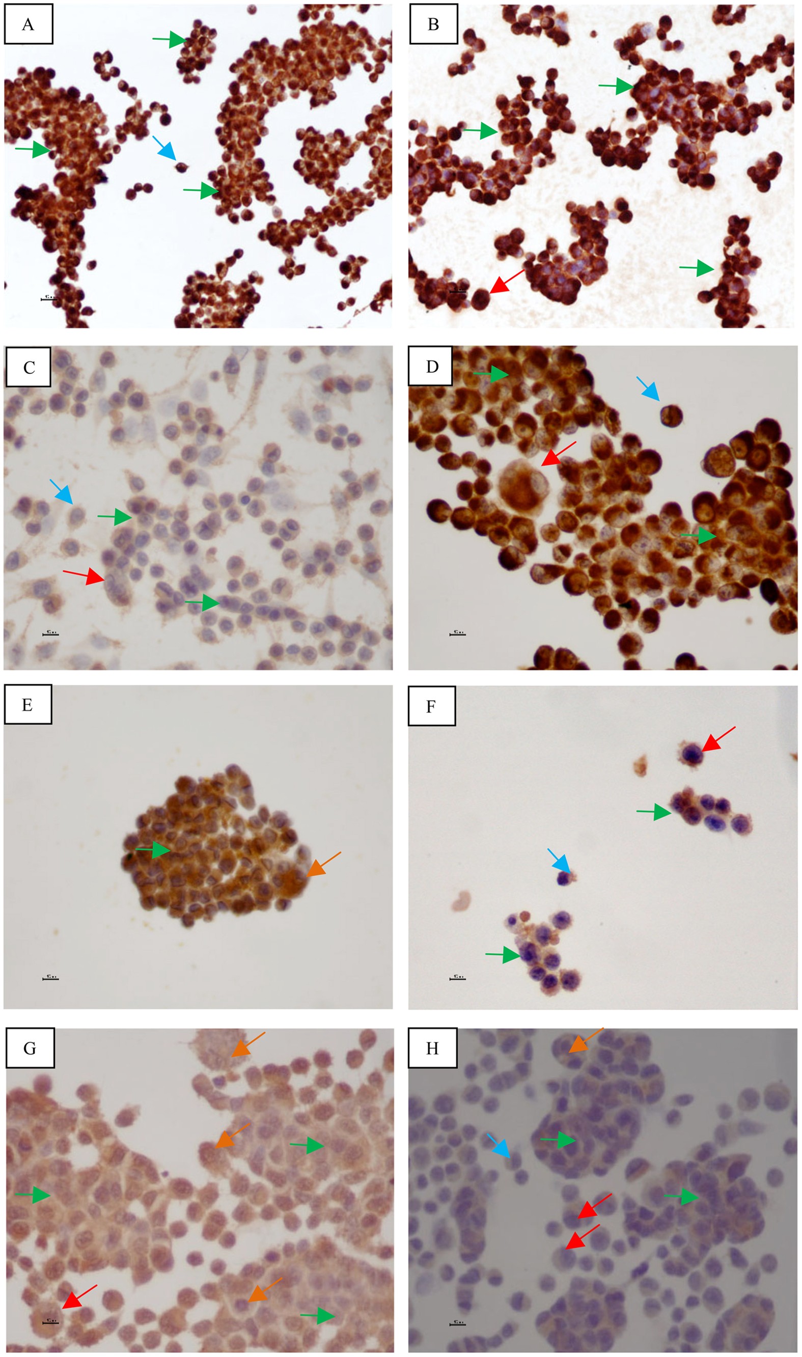
MIA PaCa-2 and PANC-1 – pancreas ductal adenocarcinoma cell lines with  neuroendocrine differentiation and somatostatin receptors | Scientific  Reports
MIA PaCa-2 and PANC-1 – pancreas ductal adenocarcinoma cell lines with neuroendocrine differentiation and somatostatin receptors | Scientific Reports
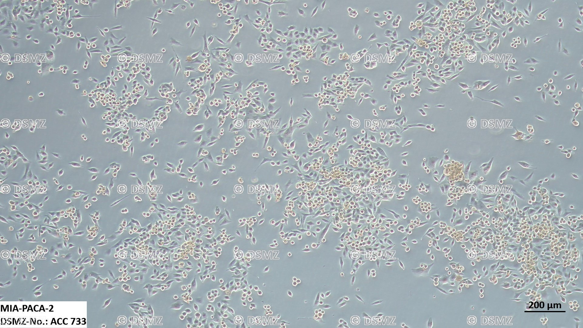
German Collection of Microorganisms and Cell Cultures GmbH: Details

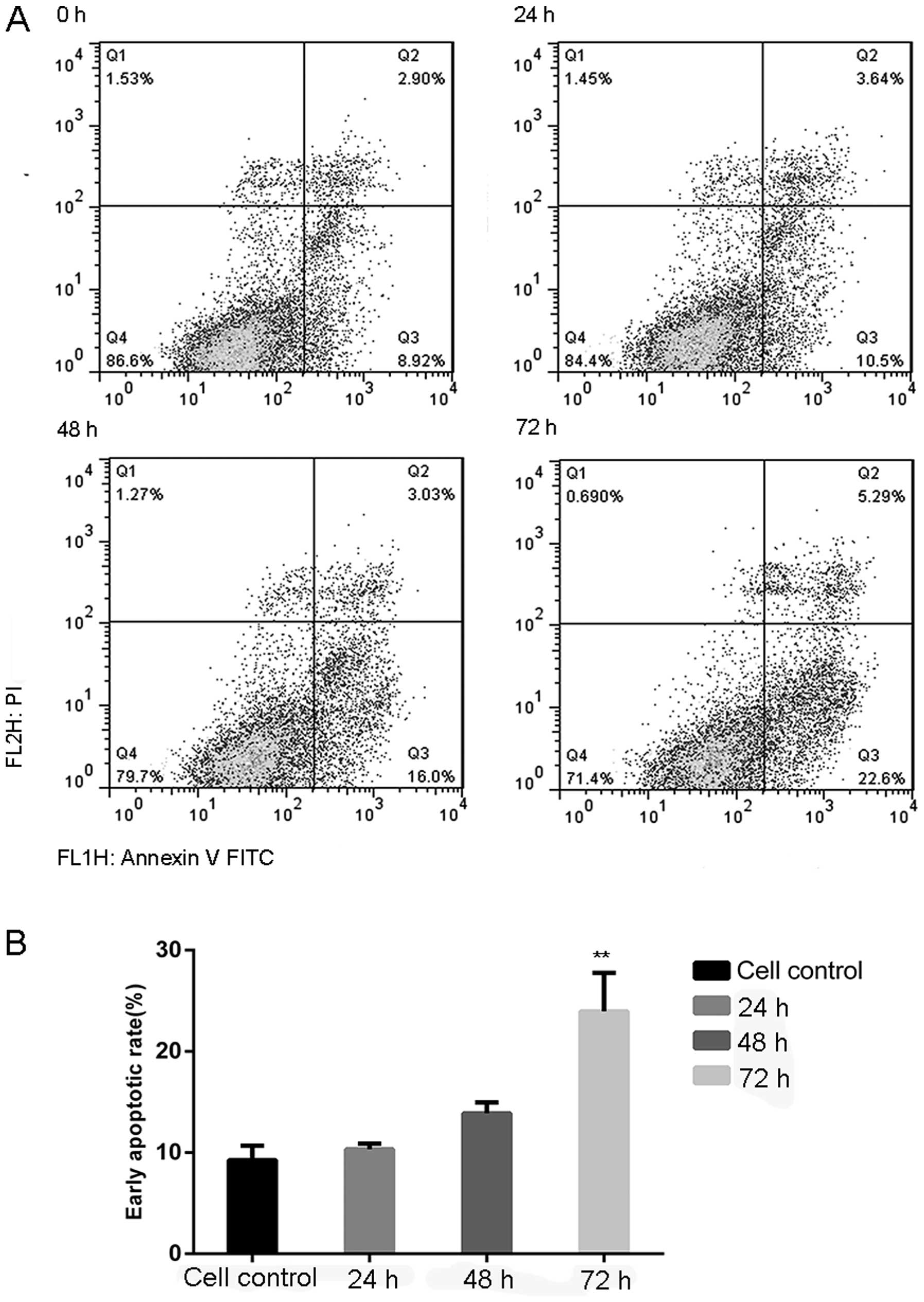
Melatonin induces cell apoptosis in Mia PaCa-2 cells via the suppression of  nuclear factor-κB and activation of ERK and JNK: A novel therapeutic  implication for pancreatic cancer
Melatonin induces cell apoptosis in Mia PaCa-2 cells via the suppression of nuclear factor-κB and activation of ERK and JNK: A novel therapeutic implication for pancreatic cancer
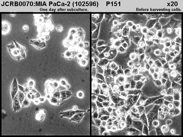
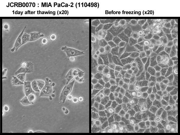
JCRB0070:MIA PaCa-2
JCRB0070:MIA PaCa-2

CELL SEARCH SYSTEM -CELL BANK- (RIKEN BRC) [RCB2094 : MIA Paca2]

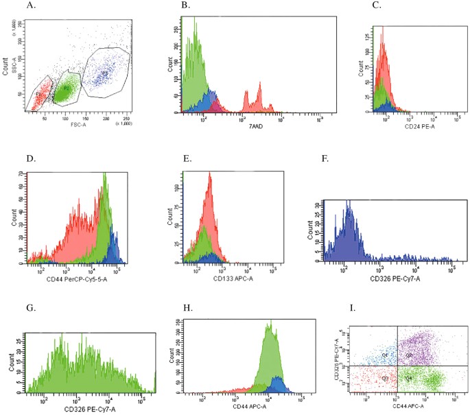
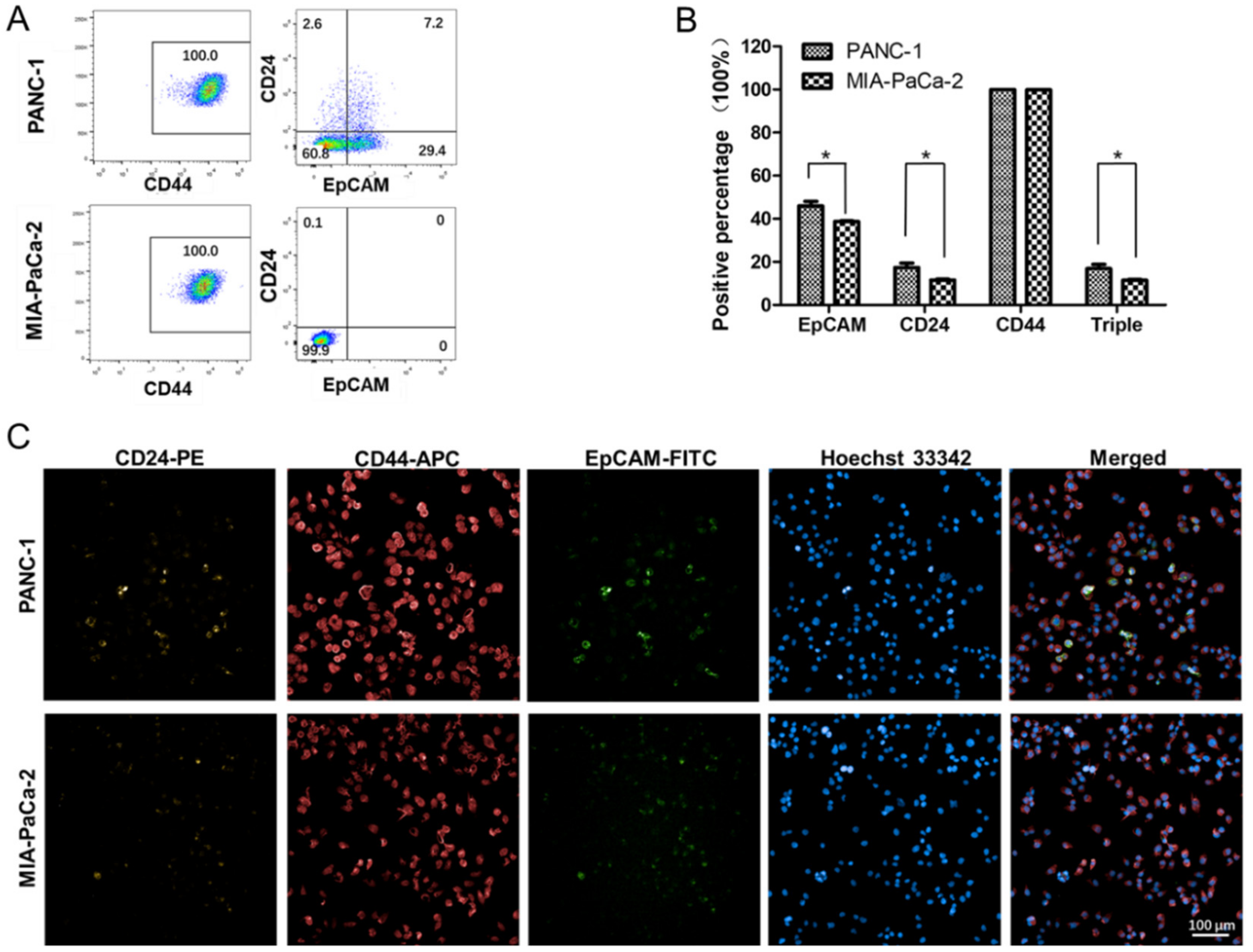
IJMS | Free Full-Text | Differentially Expressed microRNAs in MIA PaCa-2  and PANC-1 Pancreas Ductal Adenocarcinoma Cell Lines are Involved in Cancer  Stem Cell Regulation | HTML
IJMS | Free Full-Text | Differentially Expressed microRNAs in MIA PaCa-2 and PANC-1 Pancreas Ductal Adenocarcinoma Cell Lines are Involved in Cancer Stem Cell Regulation | HTML

AKT inhibition is associated with chemosensitisation in the pancreatic  cancer cell line MIA-PaCa-2 | British Journal of Cancer
AKT inhibition is associated with chemosensitisation in the pancreatic cancer cell line MIA-PaCa-2 | British Journal of Cancer

Comparison of TPPI localization in MIA PaCa-2 cells with the lysosomal... |  Download Scientific Diagram
Comparison of TPPI localization in MIA PaCa-2 cells with the lysosomal... | Download Scientific Diagram

CM03 treatment reduces tumor volume in a MIA PaCa-2 xenograft model of... |  Download Scientific Diagram
CM03 treatment reduces tumor volume in a MIA PaCa-2 xenograft model of... | Download Scientific Diagram

Gatifloxacin causes activation of p21 in MIA PaCa-2 and p27 in Panc-1  cells, but Skp2 in both cell lines.
Gatifloxacin causes activation of p21 in MIA PaCa-2 and p27 in Panc-1 cells, but Skp2 in both cell lines.
Portal do Conhecimento do Departamento de Bioquímica

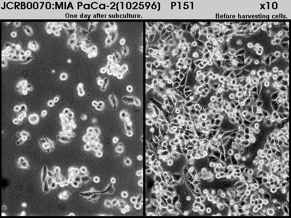
JCRB0070:MIA PaCa-2
JCRB0070:MIA PaCa-2

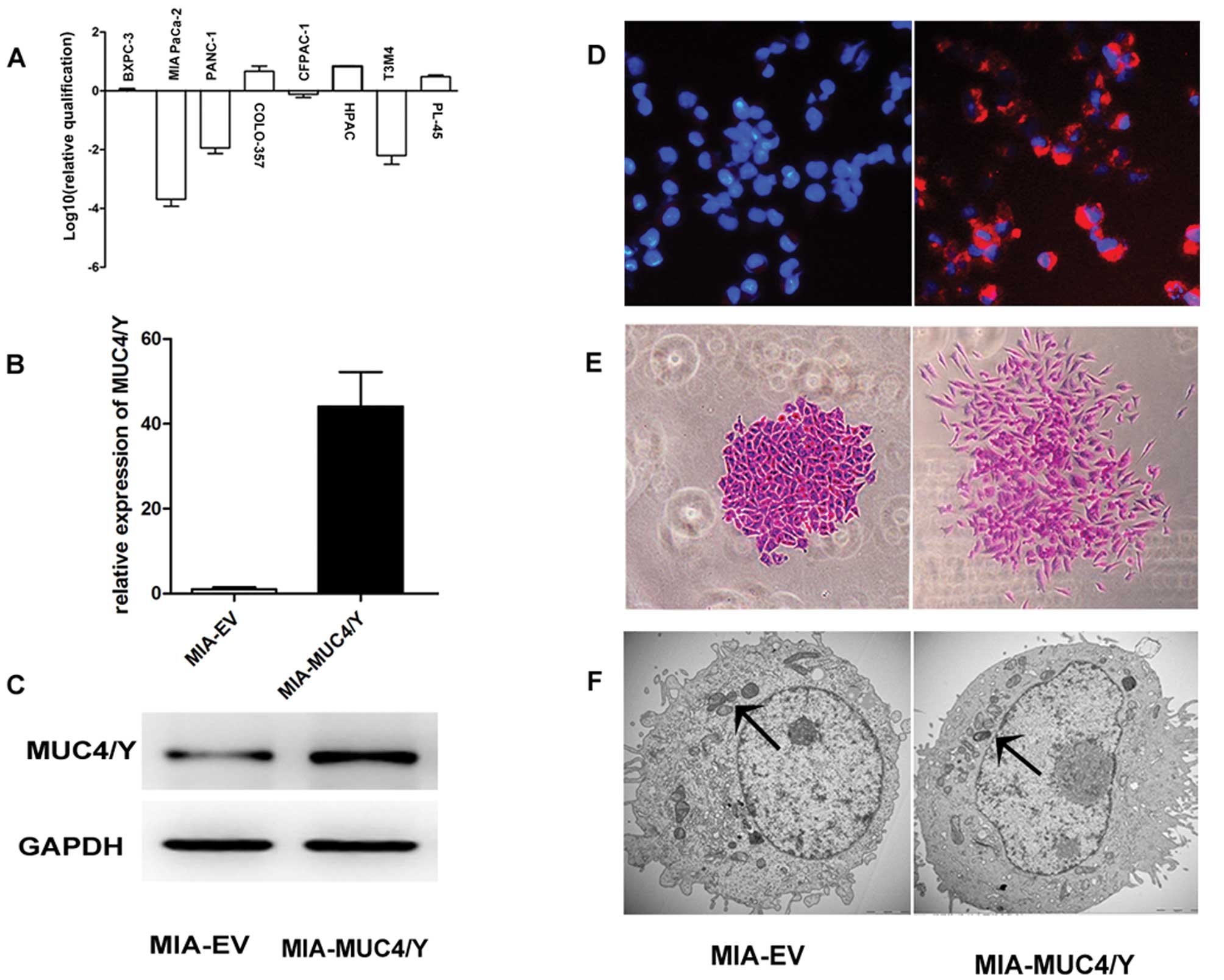
Upregulation of the splice variant MUC4/Y in the pancreatic cancer cell  line MIA PaCa-2 potentiates proliferation and suppresses apoptosis: New  insight into the presence of the transcript variant of MUC4
Upregulation of the splice variant MUC4/Y in the pancreatic cancer cell line MIA PaCa-2 potentiates proliferation and suppresses apoptosis: New insight into the presence of the transcript variant of MUC4

PLOS ONE: MicroRNA Profiling Implies New Markers of Gemcitabine  Chemoresistance in Mutant p53 Pancreatic Ductal Adenocarcinoma
PLOS ONE: MicroRNA Profiling Implies New Markers of Gemcitabine Chemoresistance in Mutant p53 Pancreatic Ductal Adenocarcinoma

Mia-PaCa-2
Mia-PaCa-2

Z-360 Suppresses Tumor Growth in MIA PaCa-2-bearing Mice via Inhibition of  Gastrin-induced Anti-Apoptotic Effects | Anticancer Research
Z-360 Suppresses Tumor Growth in MIA PaCa-2-bearing Mice via Inhibition of Gastrin-induced Anti-Apoptotic Effects | Anticancer Research

Reversion of Transcriptional Repression of Sp1 by 5 aza-2′ Deoxycytidine  Restores TGF-β Type II Receptor Expression in the Pancreatic Cancer Cell  Line MIA PaCa-2 | Cancer Research
Reversion of Transcriptional Repression of Sp1 by 5 aza-2′ Deoxycytidine Restores TGF-β Type II Receptor Expression in the Pancreatic Cancer Cell Line MIA PaCa-2 | Cancer Research

Spheroid cells in MIA PaCa-2, Panc-1, and PC-3. A, Pancreatic cancer... |  Download Scientific Diagram
Spheroid cells in MIA PaCa-2, Panc-1, and PC-3. A, Pancreatic cancer... | Download Scientific Diagram
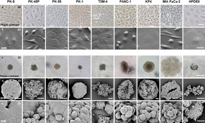
Morphofunctional analysis of human pancreatic cancer cell lines in 2- and  3-dimensional cultures | Scientific Reports
Morphofunctional analysis of human pancreatic cancer cell lines in 2- and 3-dimensional cultures | Scientific Reports
E-cadherin Stable MIA PaCa-2 Cell Line

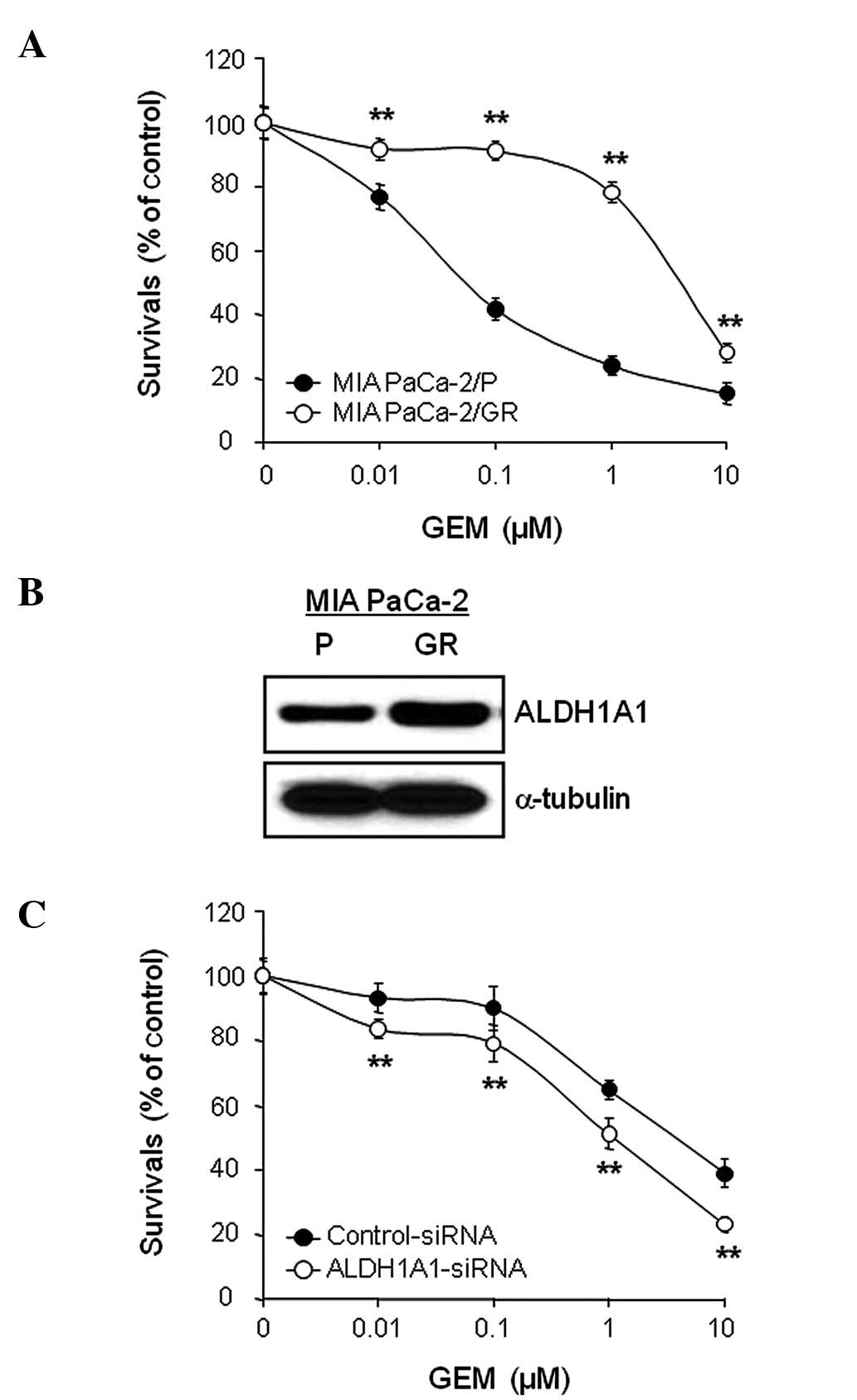
Aldehyde dehydrogenase 1A1 confers intrinsic and acquired resistance to  gemcitabine in human pancreatic adenocarcinoma MIA PaCa-2 cells
Aldehyde dehydrogenase 1A1 confers intrinsic and acquired resistance to gemcitabine in human pancreatic adenocarcinoma MIA PaCa-2 cells

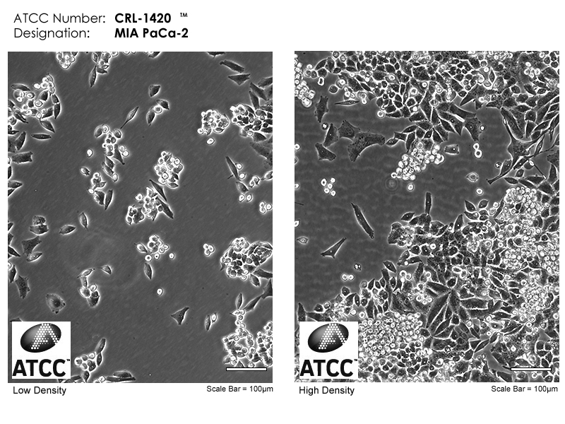
MIA PaCa-2 | ATCC
MIA PaCa-2 | ATCC

Morphological changes of MIA PaCa-2 and HPDE cells when treated with... |  Download Scientific Diagram
Morphological changes of MIA PaCa-2 and HPDE cells when treated with... | Download Scientific Diagram

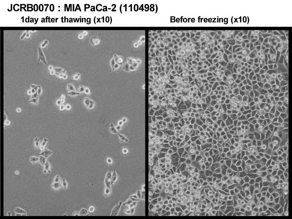
JCRB0070:MIA PaCa-2
JCRB0070:MIA PaCa-2

JCRB0070:MIA PaCa-2
JCRB0070:MIA PaCa-2

Pancreatic MIA PaCa-2 cells were stably transfected with cDNAs encoding...  | Download Scientific Diagram
Pancreatic MIA PaCa-2 cells were stably transfected with cDNAs encoding... | Download Scientific Diagram
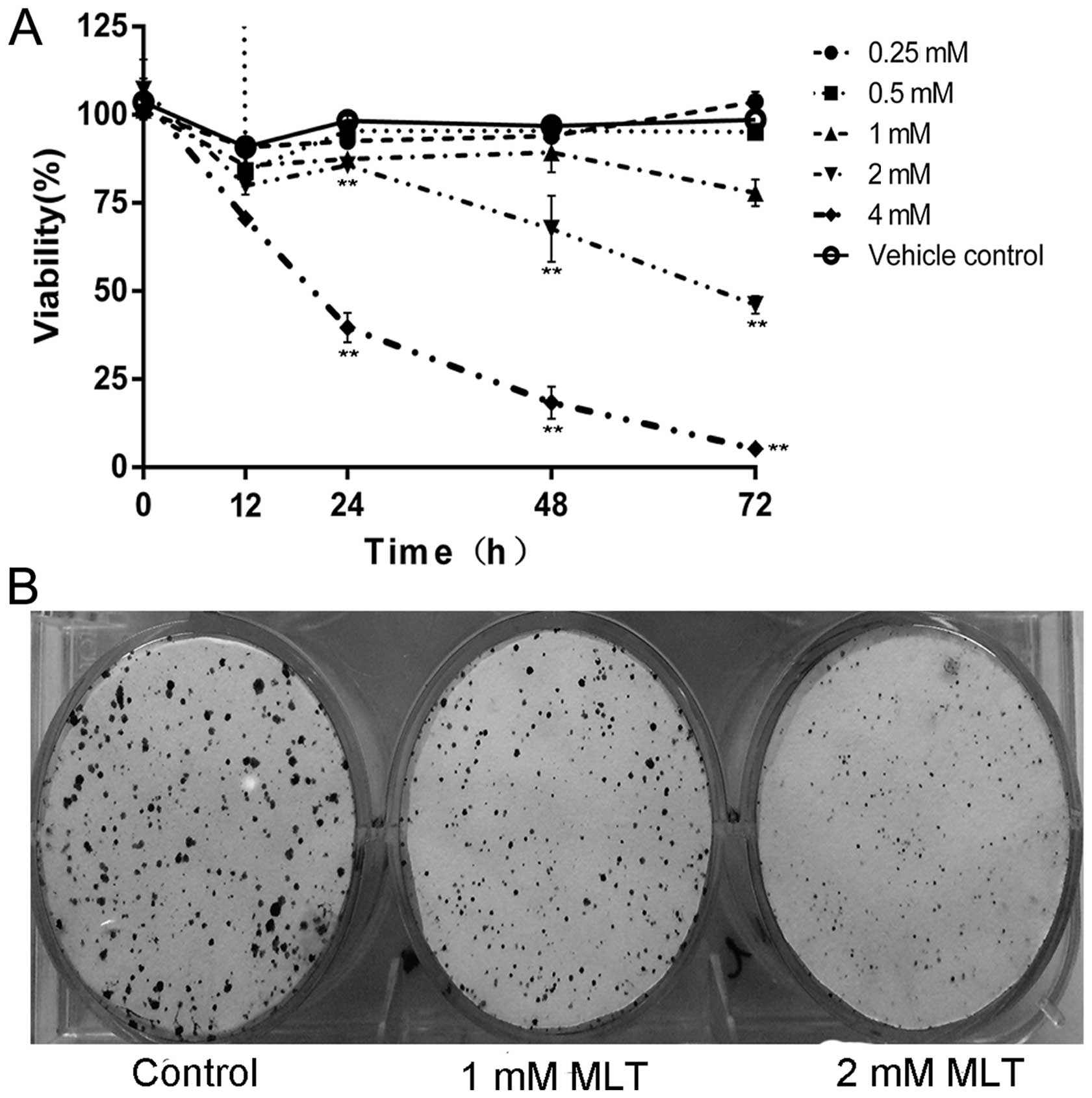
Melatonin induces cell apoptosis in Mia PaCa-2 cells via the suppression of  nuclear factor-κB and activation of ERK and JNK: A novel therapeutic  implication for pancreatic cancer
Melatonin induces cell apoptosis in Mia PaCa-2 cells via the suppression of nuclear factor-κB and activation of ERK and JNK: A novel therapeutic implication for pancreatic cancer